Maternal fetal medicine

Maternal-fetal medicine allows monitoring of pregnancies. Specific follow-up is thus provided for high-risk pregnancies, i.e., more complex cases that require specialized consultations depending on the problems presented.

What we deal with

  • Ultrasound screening examinations, fetal and maternal Doppler examinations, echocardiography, extended neurosonography, extended ultrasound diagnostics including 3D ultrasound.
  • Early diagnosis of fetal anomalies.
  • Screening for chromosomal defects.
  • Development of safertechniques for prenatal diagnosis.
  • Prediction and prevention of preeclampsia.
  • Prediction and prevention of preterm birth.
  • Prediction and prevention of stillbirth.
  • Prediction and management of fetal growth restriction.
  • Problems of multiple pregnancies.

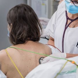
Prenatal diagnosis is a set of investigations, both instrumental and laboratory, that monitor certain aspects of the health status of the fetus during pregnancy, from the earliest stages of embryonic development to the moments before delivery.

Diagnosis and treatment

Prenatal diagnostic tests

  • First trimester test: is performed between the 11th and 13th+6th weeks of pregnancy. Based on ultrasound examinations, the hematological picture and the mother's age, the test calculates the probability of a chromosomal abnormality for the unborn child. Even in the case of a suspicious or unclear outcome, the result is still not a definite diagnosis, which is why further tests are necessary.
  • Genetic tests in pregnancy: if the result of the first trimester test is abnormal or if there is already a hereditary disease in the family, a genetic test can be used to detect certain chromosomal abnormalities or genetic diseases in the unborn child. There are two types of tests:
    • noninvasive prenatal testing (NIPT) screening for trisomies 21,13,18: a sample of the unborn child's DNA is needed, which can be extracted from the mother's blood;
    • invasive prenatal testing: a sample is taken from the placenta (villocentesis) or amniotic fluid (amniocentesis).
  • First trimester ultrasound examination, which allows:
    • accurate dating of the pregnancy;
    • early detection of major fetal abnormalities;
    • detection of multiple pregnancies with certain diagnosis of chorionicity, an essential element in the course of these pregnancies;
    • monitoring of twin pregnancies;
    • identification of women at increased risk for the development of pre-eclampsia;
    • fetal echocardiography: in-depth examination aimed at detecting possible malformations and congenital pathologies affecting the heart and great vessels.
  • Second trimester ultrasound examination: ultrasound also known as morphological ultrasound, which is performed around 20 weeks (19-21st) and has the main objective of checking the baby's size and the normality of its anatomy:
    • morphologic ultrasound performed routinely, termed Level I: if everything is normal, the woman can safely wait for the next ultrasound, in the third trimester. If, on the other hand, the examination reveals any abnormality in the fetal anatomy, the doctor will refer to an ultrasound called level II;
    • Level II ultrasonography: is distinguished by greater expertise of the operator in the area of abnormalities in the fetal anatomy. In addition, it takes place with the involvement multidisciplinary expertise (geneticists, neonatologists, etc.) that can help the couple better frame any disease diagnosis.

Treatments

  • Urgent and non-emergent medical and obstetric care for pregnant women with infectious disease: Cytomegalovirus, Hepatitis, Herpes infection, Listeria, Parvovirus B19 infection, Measles, Rubella, Syphilis, Tetanus, Toxoplasmosis, Varicella, HIV.
  • Follow-up and management ofhypertension and preeclampsia.
  • Monitoring and management of diabetes during pregnancy.
  • Monitoring and management of intrauterine growth retardation (IUGR).
  • Follow-up and management of threatened preterm delivery, cervical cerclage.
  • External maneuver version.
  • Caesarean section with high hemorrhagic risk.
  • Management of postpartumhemorrhage, multidisciplinary management, conservative surgical approach, selective embolization of pelvic vessels such as uterine and cervicovaginal arteries.

For consultations on specific cases, the EOC Maternal Fetal Medicine Service collaborates with theUniversity Hospital of Bern and the Swiss Society of Ultrasonography in Medicine (SSUM).

The time of delivery and postpartum is managed together with the obstetrics-maternity subspecialty, which offers tailored care for all mothers.

Doctors

  • Dr. med.

    Tangorra Tommaso

    Primario

Contact

Ambulatorio di Medicina materno fetale

Ospedale Regionale di Lugano, Civico

  • Orari di risposta al telefono
    Monday : 09:30 - 16:00
    Tuesday : 09:30 - 16:00
    Wednesday : 09:30 - 16:00
    Thursday : 09:30 - 16:00
    Friday : 09:30 - 16:00

  • Chiusura dell’ambulatorio
    Saturday
    Sunday

Locations

The specialty of maternal fetal medicine is part of the Department of Gynecology and Obstetrics EOC. Specialist outpatient clinics for the management of major pregnancy problems are active at Lugano Regional Hospital (Civic).

The English version of this page was created with the aid of automatic translation tools and may contain errors and omissions.
The original version is the page in Italian.